Application of microfabrication technology
to retinal prosthesis development
DB Shire, Ph.D., JL Wyatt, Ph.D., and J.F. Rizzo
III, M.D.
VA Center for Innovative Visual Rehabilitation,
Jamaica Plains, Boston, MA
Objectives: The purpose of this effort is
to develop a chronically implantable retinal prosthesis to restore useful
vision to patients who are blind with retinal diseases such as age-related
macular degeneration (AMD). AMD is the leading cause of degenerative vision
loss in the veteran population and in the developed world. There is no
known cure for this disease, and its progress can only be slowed using
currently available treatments. We are applying state-of-the-art microfabrication
technology in micro-electromechanical systems (MEMS) to develop a process
for creating the structures necessary to interface with the delicate retinal
tissue. Two examples of this effort are a model flexible, inflatable prosthesis
which is insertable through a narrow incision and may be expanded inside
the eye to assume its proper shape for implantation, and micromachined
retinal tacks which serve as a means of attaching our device to the retinal
surface with minimal trauma to nearby tissue.
Methods: In the case of the inflatable retinal
prosthesis, a central silicon hub which will contain the CMOS stimulating
circuitry is micromachined to create a nib for attachment of a flexible
silicone tube carrying compressed air to the implant. An array of flexible
polyimide 'tentacles' radiates outward from this central hub, and embedded
in each is a microchannel to facilitate inflation after insertion through
a small pars plana incision in the sclera. These structures also contain
the array of iridium oxide stimulating electrodes. In the case of the
retinal tacks, Bosch process silicon micromachining techniques have been
applied to the problem of developing a suitable means for attaching the
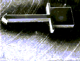
above prosthesis to the eye in a biocompatible manner. An SEM micrograph
of a sample tack is shown in the figure. Advanced photolithography techniques
have recently been used to develop sharper tack points to facilitate insertion,
and these will be demonstrated at the meeting.
Results: A model inflatable retinal prosthesis
has been developed which may be bent into a nearly arbitrary state to
facilitate insertion of the device through a narrow incision (for safety),
yet may be inflated once inside the eye to over 9 mm in diameter. This
device thus may be used to stimulate an area of the retina covering over
30 degrees of the patient's former field of view. Its flexible design
also allows the implant to conform to the spherical shape of the anterior
surface of the eye. Custom designed micromachined retinal tacks have also
been developed for the purpose of attaching this device to the retina.
Conclusions: The application of recent developments
in microfabrication technology to retinal prosthesis development is ushering
in an exciting new era in visual rehabilitation research. The present
efforts represent but two examples of the potential of collaboration between
the nanotechnology and rehabilitation R&D communities.
Funding acknowledgment: This work was funded
by the VA Rehabilitation Research and Development Service.