Local measures of psychophysical sensitivity,
visual acuity and electroretinographic function in patients with age-related
macular degeneration
J. Szlyk, Ph.D.1,2, W. Seiple, Ph.D.1,3, J. Paliga, BS2, T.S. Vajaranant, MD2, N.P. Blair, MD2, J.S. Pulido. MS, MD2
1Research
and Development Service, Chicago Veterans Administration Health Care System,
West Side Division, 2University of Illinois at Chicago, 3New
York University School of Medicine
Objectives: Our laboratory has developed
a system to measure visual acuity at 27 discrete locations. Our rationale
for the development of the instrument was to identify areas of remaining
vision to be utilized for eccentric "surrogate" fixation areas
for patients with central retinal diseases. As a validation of this new
technology, the objective of this study was to examine the relationships
among psychophysical and electroretinographic (multifocal ERG) measures
of central visual function.
Methods: Twelve patients with non-exudative
age-related macular degeneration were recruited. The patients ranged in
age from 54 to 82 years (Median = 77.5 yrs.) and visual acuities ranging
from 20/20 to 20/200. MfERG responses were recorded using 103 scaled hexagons.
Humphrey visual field thresholds were measured at locations corresponding
to the mfERG hexagons within the central 10° of the visual field. Local
visual acuity was measured using an instrument that we recently developed
that allows stimulus presentation under direct fundus viewing. This ensured
accurate placement of targets on the retina. Acuity was measured in 27
locations within the central 10°.
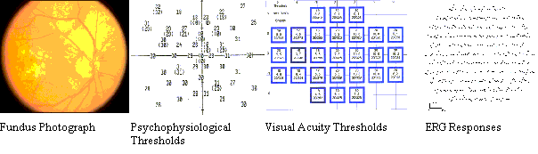 (Szlyk) A representative case
of AMD patient with geographic atrophy in the macula.
(Szlyk) A representative case
of AMD patient with geographic atrophy in the macula.
Results. The patients' mean visual acuities
were significantly worse than normal at all 27 locations tested. Visual
field thresholds were also elevated at 42 of 45 locations. Local analyses
of the mfERG show reduced amplitudes and delayed implicit times, predominantly
within the central 10°.
Conclusions: There was a good correspondence
among the three measures of central retinal function.
Funding Acknowledgment: This study was funded
by the Department of Veterans Affairs, Rehabilitation Research and Development
Service, project # C2478R.